publications
X for exploring the unknown.
main publications
#: first author; *: corresponding author.
2025
-
 Niu Liu#, Jiaying Wu#, Enze Deng#, Jianglong Zhong#, Bin Wei#, Tingting Cai#, Zhijun Xie, Xiaohui Duan, Sha Fu, David O. Osei-Hwedieh, Kezhi Huang, Peilin Zhuang, Ou Sha, Yunsheng Chen, Xiaobin Lv, Yingying Zhu, Lizao Zhang, Hsinyu Lin, Qunxing Li, Peichia Lu, Jiahao Miao, Teppei Yamada, Lei Cai, Hongwei Du, Sylvan C. Baca, Qingpei Huang, Soldano Ferrone, Xinhui Wang*, Fang Xu*, Xiaoying Fan*, and Song Fan*Nature Medicine, Aug 2025
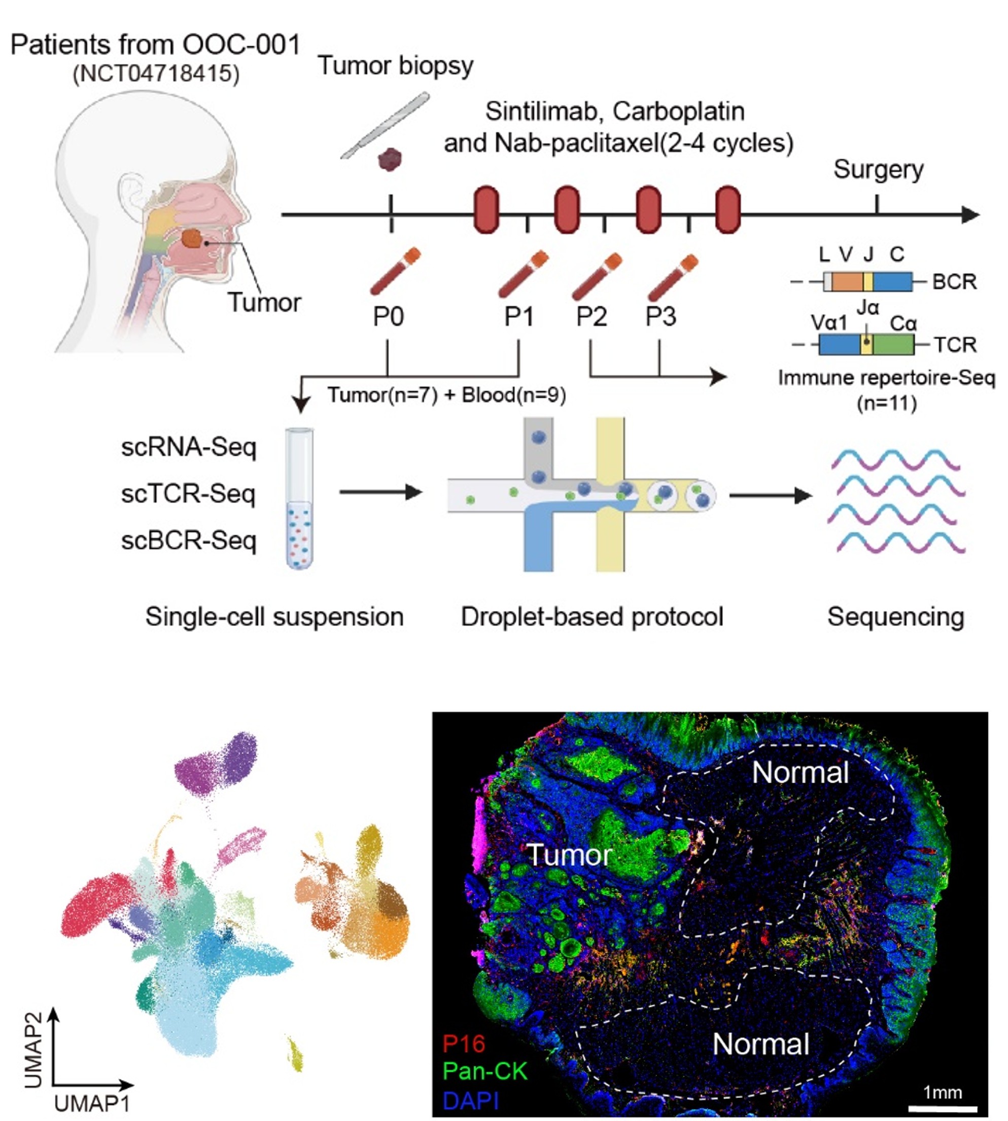
Recent advancements in cancer immunotherapy have improved patient outcomes, yet responses to immunotherapy remain moderate. Immunosenescence has been shown to contribute to the development and progression of various diseases; however, its specific role in solid tumors has not been fully delineated. Here we conducted a phase 2 clinical trial involving 51 patients with cancer undergoing neoadjuvant chemoimmunotherapy and applied single-cell RNA as well as TCR and BCR sequencing on tumor and blood samples to elucidate the immune cell perturbations. Our findings associate poor response with reduced levels of CCR7+ CD4+ naive T cells and CD27+ memory B cells, as well as higher expression of immunosenescence-related genes in T and B cell subsets. Using naturally aged mice and Ercc1-deficient mice (premature aging), we found that senolytics enhance the therapeutic efficacy of immunotherapy in multiple solid tumors by mitigating immunosenescence. Notably, we launched a phase 2 clinical trial (COIS-01) investigating the combination of senolytics with anti-PD-1 therapy. The results showed that the combination therapy achieved a 33.3% (95% confidence interval 16.6–54.7%) major pathological response rate with a low incidence of grade 3–4 adverse events (4.2%). These findings underscore the pivotal role of immunosenescence characteristics in influencing the effectiveness of immunotherapy and suggest a promising therapeutic efficacy along with a favorable safety for the combination of senolytics with anti-PD-1 therapy. ClinicalTrials.gov Identifier: OOC-001(NCT04718415) and COIS-01(NCT05724329).
To be replaced by AI response using the paper as the prompt.
-
 Siyu Li#, Yan Shen#, Yefei Chen#, Zexuan Hong, Lewei Zhang, Lufeng Ding, Chao-Yu Yang, Xiaoyang Qi, Quqing Shen, Yanyang Xiao, Pak-Ming Lau*, Zhonghua Lu*, Fang Xu*, and Guo-Qiang BiNeuroscience Bulletin, Jan 2025
To be replaced by AI response using the paper as the prompt.
2024
-
 Chao-Yu Yang#, Yan Shen, Xiaoyang Qi, Lufeng Ding, Yanyang Xiao, Qingyuan Zhu, Hao Wang, Cheng Xu, Pak-Ming Lau, Pengcheng Zhou*, Fang Xu*, and Guo-Qiang Bi*Neuroscience Bulletin, Nov 2024
To be replaced by AI response using the paper as the prompt.
2023
-
 Dong-Qing Shi#, Fang Xu#, Guo-Qiang Bi*, and Pak-Ming Lau*Neuroscience Bulletin, Nov 2023
To be replaced by AI response using the paper as the prompt.
2021
-
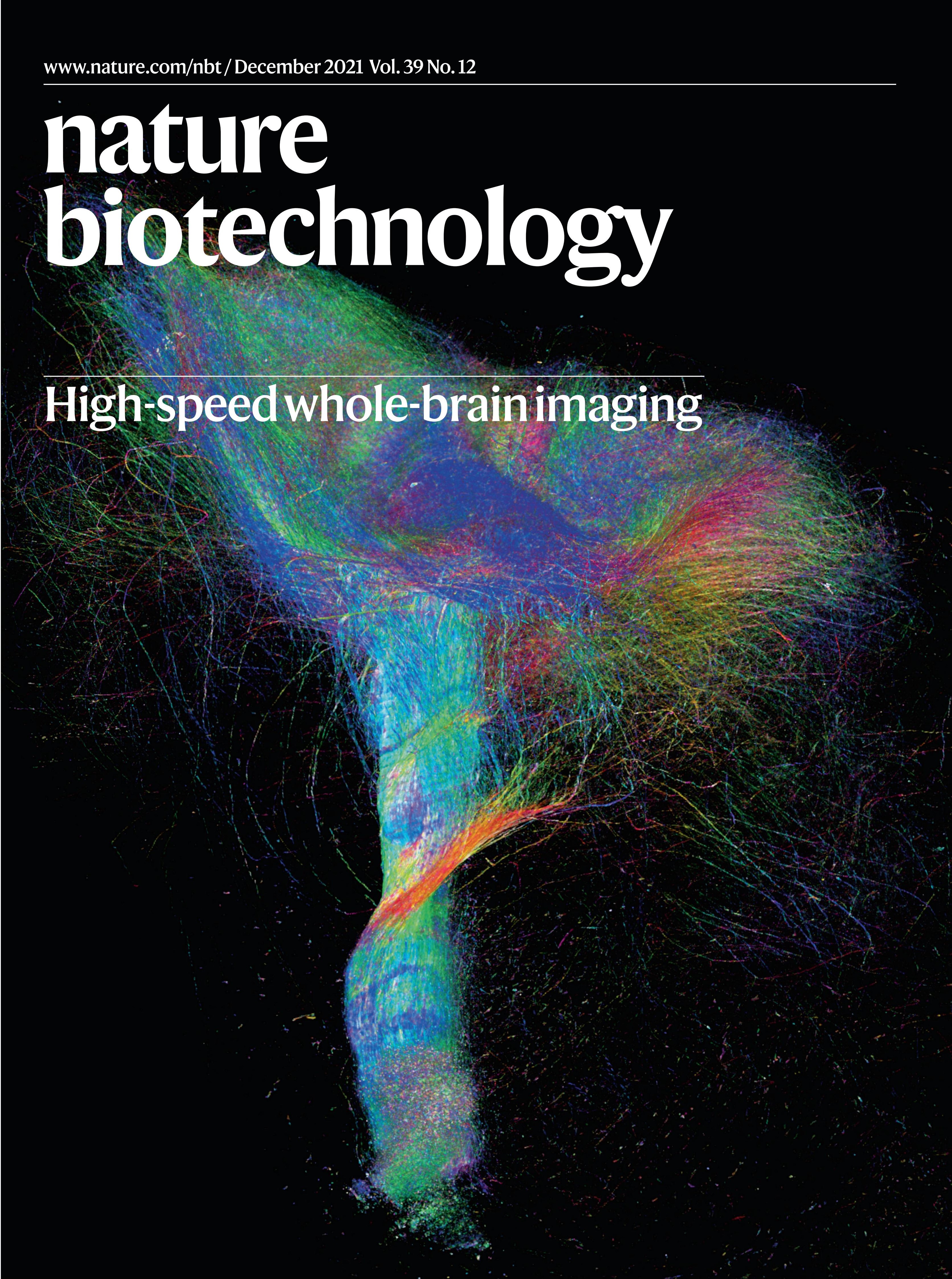 Fang Xu#, Yan Shen#, Lufeng Ding#, Chao-Yu Yang#, Heng Tan, Hao Wang, Qingyuan Zhu, Rui Xu, Fengyi Wu, Yanyang Xiao, Cheng Xu, Qianwei Li, Peng Su, Li I. Zhang, Hong-Wei Dong, Robert Desimone, Fuqiang Xu, Xintian Hu, Pak-Ming Lau*, and Guo-Qiang Bi*Nature Biotechnology, Jul 2021
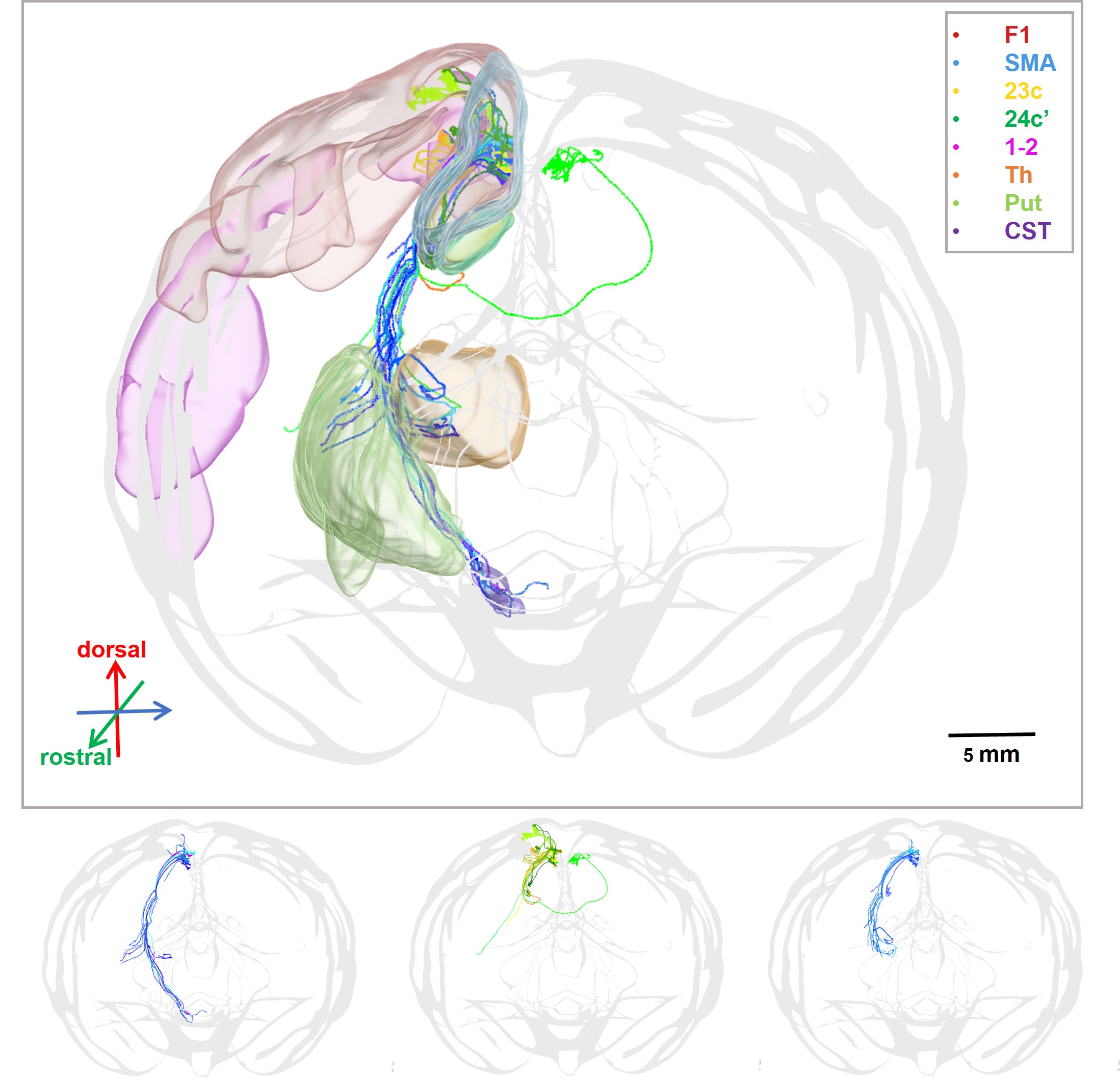
Whole-brain mesoscale mapping in primates has been hindered by large brain sizes and the relatively low throughput of available microscopy methods. Here, we present an approach that combines primate-optimized tissue sectioning and clearing with ultrahigh-speed fluorescence microscopy implementing improved volumetric imaging with synchronized on-the-fly-scan and readout technique, and is capable of completing whole-brain imaging of a rhesus monkey at 1 × 1 × 2.5 µm3 voxel resolution within 100 h. We also developed a highly efficient method for long-range tracing of sparse axonal fibers in datasets numbering hundreds of terabytes. This pipeline, which we call serial sectioning and clearing, three-dimensional microscopy with semiautomated reconstruction and tracing (SMART), enables effective connectome-scale mapping of large primate brains. With SMART, we were able to construct a cortical projection map of the mediodorsal nucleus of the thalamus and identify distinct turning and routing patterns of individual axons in the cortical folds while approaching their arborization destinations.
To be replaced by AI response using the paper as the prompt.
2018
-
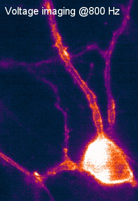 Fang Xu#, Dong-Qing Shi, Pak-Ming Lau, Michael Z. Lin*, and Guo-Qiang Bi*Molecular Brain, Jul 2018
Recent interest in high-throughput recording of neuronal activity has motivated rapid improvements in genetically encoded calcium or voltage indicators (GECIs or GEVIs) for all-optical electrophysiology. Among these probes, the ASAPs, a series of voltage indicators based on a variant of circularly permuted green fluorescent protein (cpGFP) and a conjugated voltage sensitive domain (VSD), are capable of detecting both action potentials and subthreshold neuronal activities. Here we show that the ASAPs, when excited by blue light, undergo reversible photobleaching. We find that this fluorescence loss induced by excitation with 470-nm light can be substantially reversed by low-intensity 405-nm light. We demonstrate that 405-nm and 470-nm co-illumination significantly improved brightness and thereby signal-to-noise ratios during voltage imaging compared to 470-nm illumination alone. Illumination with a single wavelength of 440-nm light also produced similar improvements. We hypothesize that reversible photobleaching is related to cis-trans isomerization and protonation of the GFP chromophore of ASAP proteins. Amino acids that influence chromophore isomerization are potential targets of point mutations for future improvements.
To be replaced by AI response using the paper as the prompt.
contributed publications
2025
- Nat.Photon.Lijuan Tang, Jiayu Wang, Jiayi Ding, Junyou Sun, Xing-jun Chen, Quqing Shen, Ruiheng Song, Peng Cao, Rong Gong, Fang Xu, Woo-ping Ge, Wenzhi Sun, Hu Zhao, and Jianglai WuNature Photonics, 2025
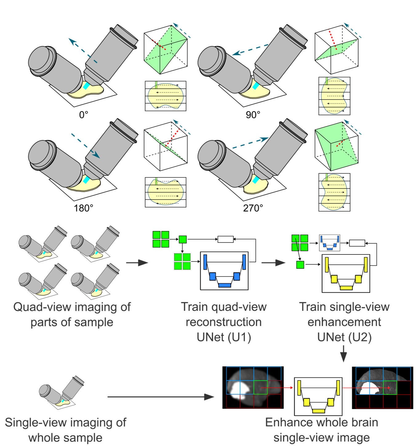
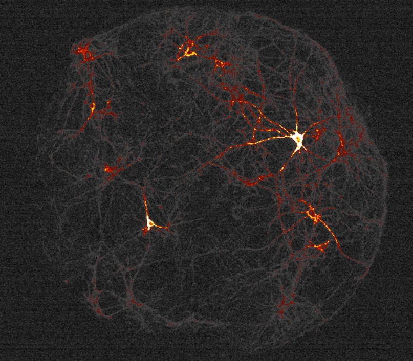
Imaging large cleared tissues requires scaling the throughput of imaging techniques. Light sheet microscopy is a promising technique for high-throughput imaging; however, its reliance on conventional microscope objectives limits the optimization of the trade-off between spatial resolution and field of view. Here we introduce curved light sheet microscope to perform optical sectioning with curved light sheets. This concept addresses the long-standing field curvature problem and lowers the barriers in designing high-throughput objectives. Leveraging a customized objective, the curved light sheet microscope achieves diffraction-limited resolution of 1.0 μm laterally and 2.5 μm axially, with uniform contrast over a field of view of more than 1 × 1 cm2. Our technique is also compatible with various tissue clearing techniques. We demonstrate that imaging an entire intact cleared mouse brain at a voxel size of 0.625 × 0.625 × 1.25 μm3 can be completed in less than 3 h, without the need for image tiling. We share a full optical description of the objective and report imaging of neuronal and vascular networks, as well as tracing of brain-wide long-distance axonal projections in intact mouse brains.
- PLoS Biol.Shun Hao, Man Xue, Qi-Yu Chen, Jinjin Wan, Yu-Jie Ma, Wantong Shi, Xuanying Chen, Xu-Hui Li, Jing-Shan Lu, Fang Xu, Guo-Qiang Bi, Wucheng Tao, and Min ZhuoPLOS Biology, 2025
The anterior cingulate cortex (ACC) is recognized as a pivotal cortical region involved in the perception of pain. The retrosplenial cortex (RSC), located posterior to the ACC, is known to play a significant role in navigation and memory processes. Although the projections from the RSC to the ACC have been found, the specifics of the synaptic connections and the functional implications of the RSC-ACC projections remain less understood. In this study, we employed a combination of whole-brain imaging, in vitro electrophysiology, and two-photon calcium imaging techniques to confirm the presence of direct excitatory glutamatergic projections from the RSC to the ACC in mice. This excitatory transmission is predominantly mediated by the postsynaptic AMPA receptors. Furthermore, the activation of the RSC-ACC projections through opto-/chemogenetics significantly facilitated the behavioral responses to both mechanical and thermal nociceptive stimuli in adult mice. Notably, this activation did not influence spinal nociceptive responses in the tail-flick test, nor did it affect anxiety-like or aversive behaviors. These findings indicate that the RSC-ACC glutamatergic pathway modulates nociceptive perception primarily at the supraspinal cortical level. We have identified a novel cortico-cortical facilitatory pathway that contributes to nociceptive processing in the cingulate cortex. The RSC-ACC pathway probably serves to integrate memory engrams with pain perception in both humans and animals.
2024
- NeuronGaowei Chen, Shishi Lai, Shaolei Jiang, Fengling Li, Kaige Sun, Xiaocong Wu, Kuikui Zhou, Yutong Liu, Xiaofei Deng, Zijun Chen, Fang Xu, Yu Xu, Kunhua Wang, Gang Cao, Fuqiang Xu, Guo-Qiang Bi, and Yingjie ZhuNeuron, 2024
The lateral septum (LS) is composed of heterogeneous cell types that are important for various motivated behaviors. However, the transcriptional profiles, spatial arrangement, function, and connectivity of these cell types have not been systematically studied. Using single-nucleus RNA sequencing, we delineated diverse genetically defined cell types in the LS that play distinct roles in reward processing. Notably, we found that estrogen receptor 1 (Esr1)-expressing neurons in the ventral LS (LSEsr1) are key drivers of reward seeking via projections to the ventral tegmental area, and these neurons play an essential role in methamphetamine (METH) reward and METH-seeking behavior. Extended exposure to METH increases the excitability of LSEsr1 neurons by upregulating hyperpolarization-activated cyclic nucleotide-gated (HCN) channels, thereby contributing to METH-induced locomotor sensitization. These insights not only elucidate the intricate molecular, circuit, and functional architecture of the septal region in reward processing but also reveal a neural pathway critical for METH reward and behavioral sensitization.
- Adv.Sci.Xu‐Hui Li, Wantong Shi, Zhi‐Xia Zhao, Takanori Matsuura, Jing‐Shan Lu, Jingmin Che, Qi‐Yu Chen, Zhaoxiang Zhou, Man Xue, Shun Hao, Fang Xu, Guo‐Qiang Bi, Bong‐Kiun Kaang, Graham L. Collingridge, and Min ZhuoAdvanced Science, 2024
The corticostriatal connection plays a crucial role in cognitive, emotional, and motor control. However, the specific roles and synaptic transmissions of corticostriatal connection are less studied, especially the corticostriatal transmission from the anterior cingulate cortex (ACC). Here, a direct glutamatergic excitatory synaptic transmission in the corticostriatal projection from the ACC is found. Kainate receptors (KAR)‐mediated synaptic transmission is increased in this corticostriatal connection both in vitro and in vivo seizure‐like activities. GluK1 containing KARs and downstream calcium‐stimulated adenylyl cyclase subtype 1 (AC1) are involved in the upregulation of KARs following seizure‐like activities. Inhibiting the activities of ACC or its corticostriatal connection significantly attenuated pentylenetetrazole (PTZ)‐induced seizure. Additionally, injection of GluK1 receptor antagonist UBP310 or the AC1 inhibitor NB001 both show antiepileptic effects. The studies provide direct evidence that KARs are involved in seizure activity in the corticostriatal connection and the KAR‐AC1 signaling pathway is a potential novel antiepileptic strategy. This study found that there is a direct glutamatergic projection from the ACC to the dorsal striatum mediated by AMPA/KA receptors. KAR‐mediated EPSCs increased in the ACC‐striatum pathway following seizure‐like activity in both in vitro and in vivo seizure models. Inhibiting the projection of ACC to the striatum significantly attenuated PTZ‐induced seizure‐like activities. The KAR‐AC1 signaling pathway thus presents a potential antiepileptic target.
2023
- Comm.Bio.Xu-Hui Li, Wantong Shi, Qi-Yu Chen, Shun Hao, Hui-Hui Miao, Zhuang Miao, Fang Xu, Guo-Qiang Bi, and Min ZhuoCommunications Biology, 2023
The brain consists of the left and right cerebral hemispheres and both are connected by callosal projections. Less is known about the basic mechanism of this cortical-cortical connection and its functional importance. Here we investigate the cortical-cortical connection between the bilateral anterior cingulate cortex (ACC) by using the classic electrophysiological and optogenetic approach. We find that there is a direct synaptic projection from one side ACC to the contralateral ACC. Glutamate is the major excitatory transmitter for bilateral ACC connection, including projections to pyramidal cells in superficial (II/III) and deep (V/VI) layers of the ACC. Both AMPA and kainate receptors contribute to synaptic transmission. Repetitive stimulation of the projection also evoked postsynaptic Ca2+ influx in contralateral ACC pyramidal neurons. Behaviorally, light activation of the ACC-ACC connection facilitated behavioral withdrawal responses to mechanical stimuli and noxious heat. In an animal model of neuropathic pain, light inhibitory of ACC-ACC connection reduces both primary and secondary hyperalgesia. Our findings provide strong direct evidence for the excitatory or facilitatory contribution of ACC-ACC connection to pain perception, and this mechanism may provide therapeutic targets for future treatment of chronic pain and related emotional disorders. A combination of slice physiology, in vivo optogenetics, and behavioral analyses shows that the cortical-cortical connection between bilateral anterior cingulate cortex facilitates pain perception and enhanced anxiety-like behaviors associated with acute and chronic pain in adult mice.
- NBXiao-Wei Li, Yi Ren, Dong-Qing Shi, Lei Qi, Fang Xu, Yanyang Xiao, Pak-Ming Lau, and Guo-Qiang BiNeuroscience Bulletin, 2023
Acetylcholine (ACh) is an important neuromodulator in various cognitive functions. However, it is unclear how ACh influences neural circuit dynamics by altering cellular properties. Here, we investigated how ACh influences reverberatory activity in cultured neuronal networks. We found that ACh suppressed the occurrence of evoked reverberation at low to moderate doses, but to a much lesser extent at high doses. Moreover, high doses of ACh caused a longer duration of evoked reverberation, and a higher occurrence of spontaneous activity. With whole-cell recording from single neurons, we found that ACh inhibited excitatory postsynaptic currents (EPSCs) while elevating neuronal firing in a dose-dependent manner. Furthermore, all ACh-induced cellular and network changes were blocked by muscarinic, but not nicotinic receptor antagonists. With computational modeling, we found that simulated changes in EPSCs and the excitability of single cells mimicking the effects of ACh indeed modulated the evoked network reverberation similar to experimental observations. Thus, ACh modulates network dynamics in a biphasic fashion, probably by inhibiting excitatory synaptic transmission and facilitating neuronal excitability through muscarinic signaling pathways.
- Cell Rep.Gaowei Chen, Shishi Lai, Guo Bao, Jincan Ke, Xiaogao Meng, Shanshan Lu, Xiaocong Wu, Hua Xu, Fengyi Wu, Yu Xu, Fang Xu, Guo-Qiang Bi, Guangdun Peng, Kuikui Zhou, and Yingjie ZhuCell Reports, 2023
The nucleus accumbens (NAc) plays an important role in motivation and reward processing. Recent studies suggest that different NAc subnuclei differentially contribute to reward-related behaviors. However, how reward is encoded in individual NAc neurons remains unclear. Using in vivo single-cell resolution calcium imaging, we find diverse patterns of reward encoding in the medial and lateral shell subdivision of the NAc (NAcMed and NAcLat, respectively). Reward consumption increases NAcLat activity but decreases NAcMed activity, albeit with high variability among neurons. The heterogeneity in reward encoding could be attributed to differences in their synaptic inputs and transcriptional profiles. Specific optogenetic activation of Nts-positive neurons in the NAcLat promotes positive reinforcement, while activation of Cartpt-positive neurons in the NAcMed induces behavior aversion. Collectively, our study shows the organizational and transcriptional differences in NAc subregions and provides a framework for future dissection of NAc subregions in physiological and pathological conditions.
- NSRFeng Xue, Fei Li, Ke-ming Zhang, Lufeng Ding, Yang Wang, Xingtao Zhao, Fang Xu, Danke Zhang, Mingzhai Sun, Pak-Ming Lau, Qingyuan Zhu, Pengcheng Zhou, and Guo-Qiang BiNational Science Review, 2023
To investigate the circuit-level neural mechanisms of behavior, simultaneous imaging of neuronal activity in multiple cortical and subcortical regions is highly desired. Miniature head-mounted microscopes offer the capability of calcium imaging in freely behaving animals. However, implanting multiple microscopes on a mouse brain remains challenging due to space constraints and the cumbersome weight of the equipment. Here, we present TINIscope, a Tightly Integrated Neuronal Imaging microscope optimized for electronic and opto-mechanical design. With its compact and lightweight design of 0.43 g, TINIscope enables unprecedented simultaneous imaging of behavior-relevant activity in up to four brain regions in mice. Proof-of-concept experiments with TINIscope recorded over 1000 neurons in four hippocampal subregions and revealed concurrent activity patterns spanning across these regions. Moreover, we explored potential multi-modal experimental designs by integrating additional modules for optogenetics, electrical stimulation or local field potential recordings. Overall, TINIscope represents a timely and indispensable tool for studying the brain-wide interregional coordination that underlies unrestrained behaviors. The world’s lightest head-mounted fluorescence microscope, weighing only 0.43 grams, has been developed and utilized to monitor neuronal activity in four deep brain regions simultaneously while the animal is freely behaving.
2022
- Nat.MethodsLei Qu, Yuanyuan Li, Peng Xie, Lijuan Liu, Yimin Wang, Jun Wu, Yu Liu, Tao Wang, Longfei Li, Kaixuan Guo, Wan Wan, Lei Ouyang, Feng Xiong, Anna C. Kolstad, Zhuhao Wu, Fang Xu, Yefeng Zheng, Hui Gong, Qingming Luo, Guoqiang Bi, Hongwei Dong, Michael Hawrylycz, Hongkui Zeng, and Hanchuan PengNature Methods, 2022
Recent whole-brain mapping projects are collecting large-scale three-dimensional images using modalities such as serial two-photon tomography, fluorescence micro-optical sectioning tomography, light-sheet fluorescence microscopy, volumetric imaging with synchronous on-the-fly scan and readout or magnetic resonance imaging. Registration of these multi-dimensional whole-brain images onto a standard atlas is essential for characterizing neuron types and constructing brain wiring diagrams. However, cross-modal image registration is challenging due to intrinsic variations of brain anatomy and artifacts resulting from different sample preparation methods and imaging modalities. We introduce a cross-modal registration method, mBrainAligner, which uses coherent landmark mapping and deep neural networks to align whole mouse brain images to the standard Allen Common Coordinate Framework atlas. We build a brain atlas for the fluorescence micro-optical sectioning tomography modality to facilitate single-cell mapping, and used our method to generate a whole-brain map of three-dimensional single-neuron morphology and neuron cell types. mBrainAligner is a cross-modal registration platform for whole mouse brains imaged with different modalities. In addition, a fluorescence micro-optical sectioning tomography-based mouse brain atlas has been generated.
- Yan Shen, Lu-Feng Ding, Chao-Yu Yang, Fang Xu, Pak-Ming Lau, and Guo-Qiang BiZoological Research, 2022
A: Schematic of VISoR microscope performing automated 3D imaging. B: SMART pipeline of serial sectioning and clearing, 3D microscopy, and semi-automated reconstruction and tracing. C: Axonal projections in macaque brain. Scale bar: 5 mm.
- Man Xue, Wan-Tong Shi, Si-Bo Zhou, Ya-Nan Li, Feng-Yi Wu, Qi-Yu Chen, Ren-Hao Liu, Zhao-Xiang Zhou, Yu-Xiang Zhang, Yu-Xin Chen, Fang Xu, Guo-Qiang Bi, Xu-Hui Li, Jing-Shan Lu, and Min ZhuoMolecular Pain, 2022
The anterior cingulate cortex (ACC) is located in the frontal part of the cingulate cortex, and plays important roles in pain perception and emotion. The thalamocortical pathway is the major sensory input to the ACC. Previous studies have show that several different thalamic nuclei receive projection fibers from spinothalamic tract, that in turn send efferents to the ACC by using neural tracers and optical imaging methods. Most of these studies were performed in monkeys, cats, and rats, few studies were reported systematically in adult mice. Adult mice, especially genetically modified mice, have provided molecular and synaptic mechanisms for cortical plasticity and modulation in the ACC. In the present study, we utilized rabies virus-based retrograde tracing system to map thalamic-anterior cingulate monosynaptic inputs in adult mice. We also combined with a new high-throughput VISoR imaging technique to generate a three-dimensional whole-brain reconstruction, especially the thalamus. We found that cortical neurons in the ACC received direct projections from different sub-nuclei in the thalamus, including the anterior, ventral, medial, lateral, midline, and intralaminar thalamic nuclei. These findings provide key anatomic evidences for the connection between the thalamus and ACC.
- Qiao-Qiong Liu, Yu-Xiao Cheng, Qi Jing, Ke-Ming Zhang, Lu-Feng Ding, Xiao-Wei Fan, Chun-Hui Jia, Fang Xu, Guo-Qiang Bi, and Pak-Ming LauMolecular Brain, 2022
The pedunculopontine nucleus (PPN) is a heterogeneous midbrain structure involved in various brain functions, such as motor control, learning, reward, and sleep. Previous studies using conventional tracers have shown that the PPN receives extensive afferent inputs from various cortical areas. To examine how these cortical axons make collateral projections to other subcortical areas, we used a dual-viral injection strategy to sparsely label PPN-targeting cortical pyramidal neurons in CaMKIIα-Cre transgenic mice. Using a high-speed volumetric imaging with on-the-fly-scan and Readout (VISoR) technique, we visualized brain-wide axonal projections of individual PPN-targeting neurons from several cortical areas, including the prelimbic region (PL), anterior cingulate area (ACA) and secondary motor cortex (MOs). We found that each PPN-projecting neuron had a unique profile of collateralization, with some subcortical areas being preferential targets. In particular, PPN-projecting neurons from all three traced cortical areas exhibited common preferential collateralization to several nuclei, with most neurons targeting the striatum (STR), lateral hypothalamic area (LHA) and periaqueductal gray (PAG), and a substantial portion of neurons also targeting the zona incerta (ZI), median raphe nucleus (MRN) and substantia nigra pars reticulata (SNr). Meanwhile, very specific collateralization patterns were found for other nuclei, including the intermediate reticular nucleus (IRN), parvicellular reticular nucleus (PARN) and gigantocellular reticular nucleus (GRN), which receive collateral inputs almost exclusively from the MOs. These observations provide potential anatomical mechanisms for cortical neurons to coordinate the PPN with other subcortical areas in performing different physiological functions.
- Wantong Shi, Man Xue, Fengyi Wu, Kexin Fan, Qi-Yu Chen, Fang Xu, Xu-Hui Li, Guo-Qiang Bi, Jing-Shan Lu, and Min ZhuoMolecular Pain, 2022
The anterior cingulate cortex (ACC) is a key cortical region that plays an important role in pain perception and emotional functions. Previous studies of the ACC projections have been collected primarily from monkeys, rabbits and rats. Due to technological advances, such as gene manipulation, recent progress has been made in our understanding of the molecular and cellular mechanisms of the ACC-related chronic pain and emotion is mainly obtained from adult mice. Few anatomic studies have examined the whole-brain projections of the ACC in adult mice. In the present study, we examined the continuous axonal outputs of the ACC in the whole brain of adult male mice. We used the virus anterograde tracing technique and an ultrahigh-speed imaging method of Volumetric Imaging with Synchronized on-the-fly-scan and Readout (VISoR). We created a three-dimensional (3D) reconstruction of mouse brains. We found that the ACC projected ipsilaterally primarily to the caudate putamen (CPu), ventral thalamic nucleus, zona incerta (ZI), periaqueductal gray (PAG), superior colliculus (SC), interpolar spinal trigeminal nucleus (Sp5I), and dorsal medullary reticular nucleus (MdD). The ACC also projected to contralateral brain regions, including the ACC, reuniens thalamic nucleus (Re), PAG, Sp5I, and MdD. Our results provide a whole-brain mapping of efferent projections from the ACC in adult male mice, and these findings are critical for future studies of the molecular and synaptic mechanisms of the ACC and its related network in mouse models of brain diseases.
2019
- Zhi-Qin John Xu, Fang Xu, Guoqiang Bi, Douglas Zhou, and David CaiEurophysics Letters, 2019
The maximum entropy principle (MEP) has been applied to study various problems in equilibrium and nonequilibrium systems in physics and other disciplines. Through analyses of numerical and experimental data, we demonstrate that the widely used entropic criteria, an assessment of the validity of MEP, can be misleading indexes as they can often fail to reflect the important difference between the observed and the MEP predicted statistical distribution. Our work demonstrates the importance of high-order statistical structures that cannot be captured by the entropic criteria and provides a cautionary tale of over-interpretation of results of MEP. This paper is dedicated to David Cai.
- NSRHao Wang, Qingyuan Zhu, Lufeng Ding, Yan Shen, Chao-Yu Yang, Fang Xu, Chang Shu, Yujie Guo, Zhiwei Xiong, Qinghong Shan, Fan Jia, Peng Su, Qian-Ru Yang, Bing Li, Yuxiao Cheng, Xiaobin He, Xi Chen, Feng Wu, Jiang-Ning Zhou, Fuqiang Xu, Hua Han, Pak-Ming Lau, and Guo-Qiang BiNational Science Review, 2019
The speed of high-resolution optical imaging has been a rate-limiting factor for meso-scale mapping of brain structures and functional circuits, which is of fundamental importance for neuroscience research. Here, we describe a new microscopy method of Volumetric Imaging with Synchronized on-the-fly-scan and Readout (VISoR) for high-throughput, high-quality brain mapping. Combining synchronized scanning beam illumination and oblique imaging over cleared tissue sections in smooth motion, the VISoR system effectively eliminates motion blur to obtain undistorted images. By continuously imaging moving samples without stopping, the system achieves high-speed 3D image acquisition of an entire mouse brain within 1.5 hours, at a resolution capable of visualizing synaptic spines. A pipeline is developed for sample preparation, imaging, 3D image reconstruction and quantification. Our approach is compatible with immunofluorescence methods, enabling flexible cell-type specific brain mapping and is readily scalable for large biological samples such as primate brains. Using this system, we examined behaviorally relevant whole-brain neuronal activation in 16 c-Fos-shEGFP mice under resting or forced swimming conditions. Our results indicate the involvement of multiple subcortical areas in stress response. Intriguingly, neuronal activation in these areas exhibits striking individual variability among different animals, suggesting the necessity of sufficient cohort size for such studies.
2018
- IEEE TNNLSXu Shen, Xinmei Tian, Tongliang Liu, Fang Xu, and Dacheng TaoIEEE Transactions on Neural Networks and Learning Systems, 2018
Dropout has been proven to be an effective algorithm for training robust deep networks because of its ability to prevent overfitting by avoiding the co-adaptation of feature detectors. Current explanations of dropout include bagging, naive Bayes, regularization, and sex in evolution. According to the activation patterns of neurons in the human brain, when faced with different situations, the firing rates of neurons are random and continuous, not binary as current dropout does. Inspired by this phenomenon, we extend the traditional binary dropout to continuous dropout. On the one hand, continuous dropout is considerably closer to the activation characteristics of neurons in the human brain than traditional binary dropout. On the other hand, we demonstrate that continuous dropout has the property of avoiding the co-adaptation of feature detectors, which suggests that we can extract more independent feature detectors for model averaging in the test stage. We introduce the proposed continuous dropout to a feedforward neural network and comprehensively compare it with binary dropout, adaptive dropout, and DropConnect on Modified National Institute of Standards and Technology, Canadian Institute for Advanced Research-10, Street View House Numbers, NORB, and ImageNet large scale visual recognition competition-12. Thorough experiments demonstrate that our method performs better in preventing the co-adaptation of feature detectors and improves test performance.
2013
- Richard C Gerkin, David W Nauen, Fang Xu, and Guo-Qiang BiMolecular Brain, 2013
During development both Hebbian and homeostatic mechanisms regulate synaptic efficacy, usually working in opposite directions in response to neuronal activity. Homeostatic plasticity has often been investigated by assaying changes in spontaneous synaptic transmission resulting from chronic circuit inactivation. However, effects of inactivation on evoked transmission have been less frequently reported. Importantly, contributions from the effects of circuit inactivation and reactivation on synaptic efficacy have not been individuated. Here we show for developing hippocampal neurons in primary culture that chronic inactivation with TTX results in increased mean amplitude of miniature synaptic currents (mEPSCs), but not evoked synaptic currents (eEPSCs). However, changes in quantal properties of transmission, partially reflected in mEPSCs, accurately predicted higher-order statistical properties of eEPSCs. The classical prediction of homeostasis – increased strength of evoked transmission – was realized after explicit circuit reactivation, in the form of cells’ pairwise connection probability. In contrast, distributions of eEPSC amplitudes for control and inactivated-then-reactivated groups matched throughout. Homeostatic up-regulation of evoked synaptic transmission in developing hippocampal neurons in primary culture requires both the inactivation and reactivation stages, leading to a net increase in functional circuit connectivity.